The eye is the organ that helps us see. It features several components which work in conjunction to give us sight. This includes the cornea, iris, pupil, lens, retina, macula, optic nerve, choroid and vitreous.
For a full breakdown of the human eye, read below.
What is the anatomy of the eye and its function?
The anatomy of the human eye provides us with the ability to see colour, differentiate between light and dark (light perception) and judge the distance between two objects (depth perception). The components of the human eye are split into different parts that work together to help us see. There are three layers of the human eye:
- Outermost layer – this is made up of the cornea and sclera
- Middle layer – this is where the choroid, ciliary body and iris are located
- Innermost layer – the retina can be found here
Parts of the human eye and their functions
Each part of the eye performs a different function that contributes to our vision:
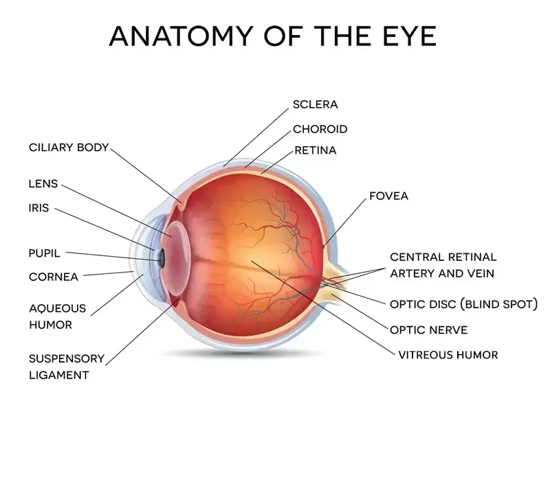
Iris – the coloured part of the eye that surrounds the pupil. It regulates the amount of light entering the eye by controlling the size of the pupil.
Pupil – the round centre (within the iris) which light passes through to focus on the retina and begin the process of sight.
Cornea – the clear part at the front of the eye which bends light rays so they can freely pass through the pupil. It also provides a physical barrier against trauma.
Lens – this is located behind the pupil and allows the eyes to focus on small details. Its main function is to transmit and focus light onto the retina.
Macula - this is located in the retina and is a small central area that contains light-sensitive cells which allow us to see in fine detail (20/20 vision).
Optic nerve – this is an extension of the central nervous system (brain and spinal cord). It transmits electrical impulses from your eyes to your brain.
Retina – located at the back of the eye, the retina contains light-sensitive cells. It converts light into chemical and nervous signals. The brain uses these signals to understand what the eye is seeing.
Choroid – this is the middle layer of the eye which is located between the retina and sclera. It contains connective tissues and supplies nutrients to the outer retina.
Ciliary body - this part of the eye connects the choroid to the iris. It produces a fluid called aqueous humor.
Fovea – the fovea (centralis) is responsible for providing the highest visual acuity, producing the sharpest vision and the greatest colour contrast.
Optic disc – also referred to as the ‘blind spot’ or optic nerve head. The optic disc is a circular area at the back of the eye where the optic nerve connects to the retina.
Sclera – the white outer layer of the eye surrounding the cornea. It supports the eyeball and protects it against rupture.
Rod cells – photoreceptor cells located in the retina at the outer edges. These cells can function in low light and are responsible for peripheral vision.
Cone cells – also located in the retina, these light-sensitive cells function best in bright light and are responsible for colour vision.
Vitreous humor – this is a transparent jelly-like substance which fills the eyeball behind the lens.
Aqueous humor – this is a thin, transparent fluid in the front part of the eye which provides nourishment to the cornea and the lens. This is 99.9% water.
How does the eye work?
The eye is a very complex organ, however, the way it works is often compared to a camera. Here’s the step-by-step process:
1) Light enters the eye through the cornea.
2) The light passes through the pupil and the iris controls the amount (of light) which passes through.
3) Light rays then reach the lens, and the rays begin to convergelens helps the eye focus on objects.
<
4) The lens focusses light rays onto the retina which receives an inverted image and converts this into electrical signals.
5) The electrical signals are transmitted by the optic nerve to the visual cortex (the part of the brain which controls sight).
Eye muscles and movement
In addition to the anatomy of the eye, there are six extraocular muscles which control eye movement:
- Lateral rectus – this pulls the pupil away from the midline of the body.
- Medial rectus – this brings the pupil closer to the midline of the body.
- Inferior rectus – this has multiple functions, the main being to help extort the eye.
- Superior rectus – this elevates the eye so the cornea can move superiorly.
- Inferior oblique – this moves the eye upwards when looking in towards the nose.
- Superior oblique – this muscle turns the eye inward to direct the gaze.
Additionally, there are different types of eye movements which are controlled by the six muscles in the eye. This includes:
- Rapid eye movement (REM) – this refers to rapid movement of the eye that happens during sleep.
- Saccade – this is controlled by the frontal lobe of the brain and refers to rapid movement of both eyes at the same time.
- Vestibulo-ocular reflex – this movement stabilises your gaze when you move your head.
- Pursuit movement – these are small tracking movements which allow you to follow a moving object.
Disclaimer: The advice in this article is for informational purposes only and does not replace medical care or an in-person check-up. Please check with an eyecare professional before purchasing any products or remedies. For information on our article review process, please refer to our Editorial Policy.


 Offers
 Account
 Menu
Menu Favorite
 Basket
 OFFERS

 Sunglasses